
Emergency Surgical Procedures
The emergency surgical procedures we perform include but are not limited to:
Soft Tissue
Colic
Generically, this refers to the clinical signs(pawing, flank watching, rolling, stretching to urinate, flehmen response) of any type of abdominal pain. The causes and severity of the colic signs seen are varied and range from simple impactions with mild signs of pain to twisted or entrapped segments of the intestinal tract that cause violent thrashing of the horse. Many types of colic can be treated with medical therapy such as stomach lavage and oral laxatives a, IV fluids and pain medications, while others require surgical intervention via an exploratory laparotomy under general anesthesia to delineate the cause and surgical treatment necessary to correct the problem.
Lacerations
Refers to a cut or tear in the skin, muscle, tendons or ligaments. Depending on the area involved and the type of injury, many can be cleaned and sutured closed while others must be left to heal via second intention (from inside out). Vital structures in some areas must be closely evaluated to determine if sepsis has developed.
Septic Joints/Tendon Sheaths
Contamination of a joint with bacteria or fungi secondary to a puncture, laceration, injection or blood borne route. This frequently requires a combination of surgical lavage, local and systemic antibiotics or anti-fungals and pain medications.
C-Section
Surgical delivery of a foal via an abdominal incision either electively or as an emergency procedure during a dystocia.
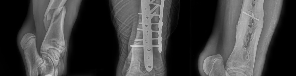
Orthopedic Surgery
Complex Fracture Repair
Routine (e.g. condylar, carpal/tarsal slab and proximal phalanx fractures, etc.)
Salter-Harris and rib fractures (foals)
Arthroscopically-guided articular fracture repair
Major Fracture
State-of-the-art locking compression bone plate technology
We use only Synthes-DePuy orthopedic implants
- Breakdown injury
- Arthrodesis (joint fusion)
Laparoscopic Procedures
Post-Castration Hemorrhage
Bleeding following castration is not an uncommon complication but has life-threatening implications, especially if the bleeding cannot be controlled. In some cases, with a standing approach, a small camera may be introduced through the body wall into the abdomen to locate the blood vessel that is the source of the hemorrhage and, using a Ligasure device, the vascular pedicle that once supplied the testicle is cauterized to stop the hemorrhage.
Diaphragmatic Hernia
A hole in the diaphragm (the musculotendinous tissue that separates the abdominal cavity from the thoracic cavity) can create serious problems, including hemorrhage or herniation of intestines into the chest cavity. By introducing a small camera into either the thoracic cavity or the abdominal cavity, the defect can be visualized and subsequently repaired.
Tension Pneumothorax
Increased pressure within the pleural space (the space between the lungs and the chest well) causes severely compromised respiration and is life-threatening. By using thorascopy and introducing a small camera into the thoracic cavity, the source of the tension pneumothorax can be elucidated and allow for relief of the intrapleural pressure.
Rectal Tear
Partial or complete perforation of the rectum is a life-threatening condition in horses that can result in an uncontrollable infection within the abdominal cavity. With sedation, local anesthesia, and epidural anesthesia, emergency repair of the defect can be performed in a standing horse. A small camera is introduced into the abdomen to allow repair of the defect with suture and/or placement of a liner to bypass the tear.